-
Recognize cell organelles, which are visible by regular light microscopy (Nucleus, nucleolus, basophilic rough endoplasmic reticulum, secretory vesicles) and by EM (Golgi complex, lysosomes, rough and smooth ER and others).
- Know different functions that are associated with different types of eukaryotic cell organelles.
- Review the secretory pathway and the function and structure of the organelles involved.
- Compare the three different cytoskeletal systems
- Know the different types of cell-cell and cell-substrate adhesion complexes and apical specializations (microvilli and cilia) and their association with cytoskeletal elements.
- Being able to recognize various cell organelles and cell junctions in electron micrographs and being able to identify striated or brush borders and cilia by light microscopy.
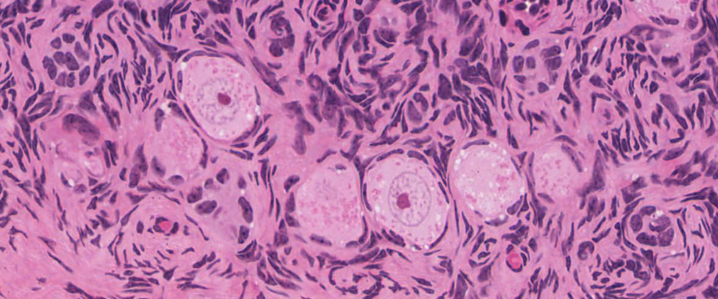
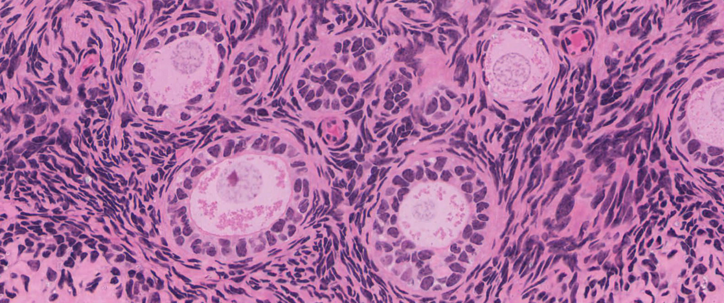
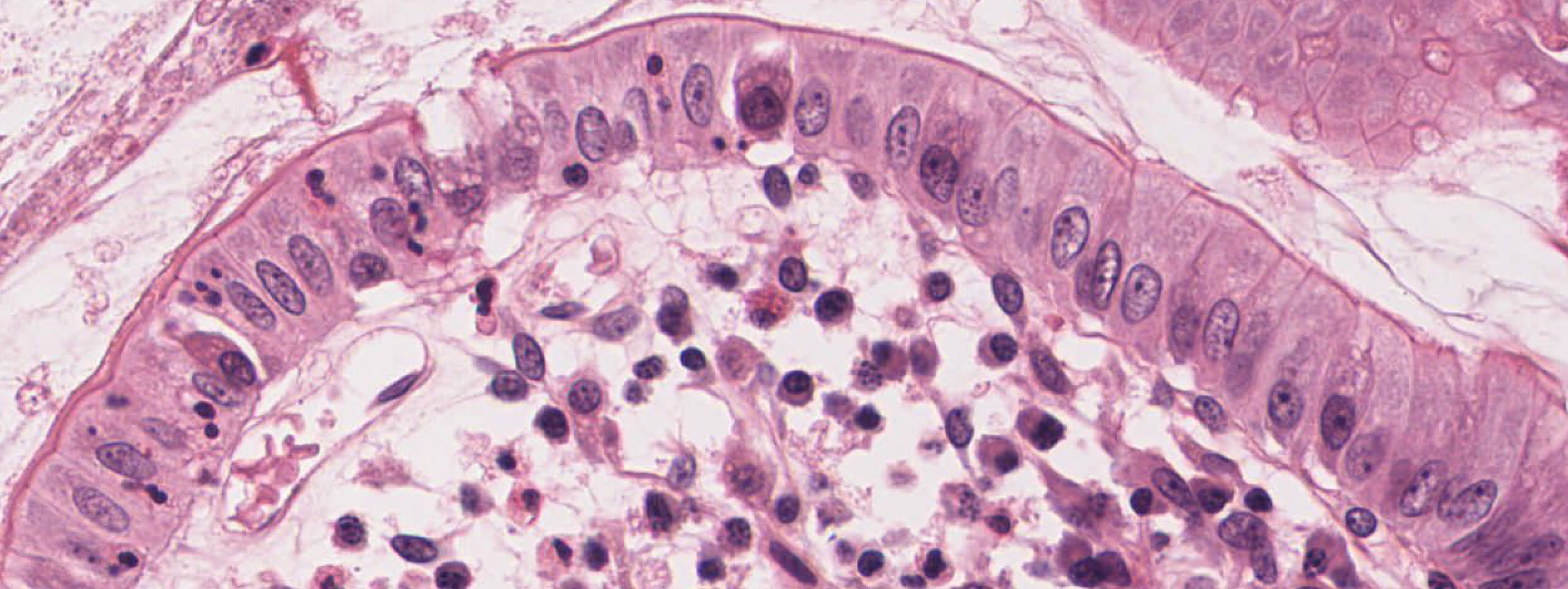
Slide 239 Ovary, monkey H&E View Virtual Slide
Slide 065-1 Spinal cord, cross section Masson View Virtual Slide
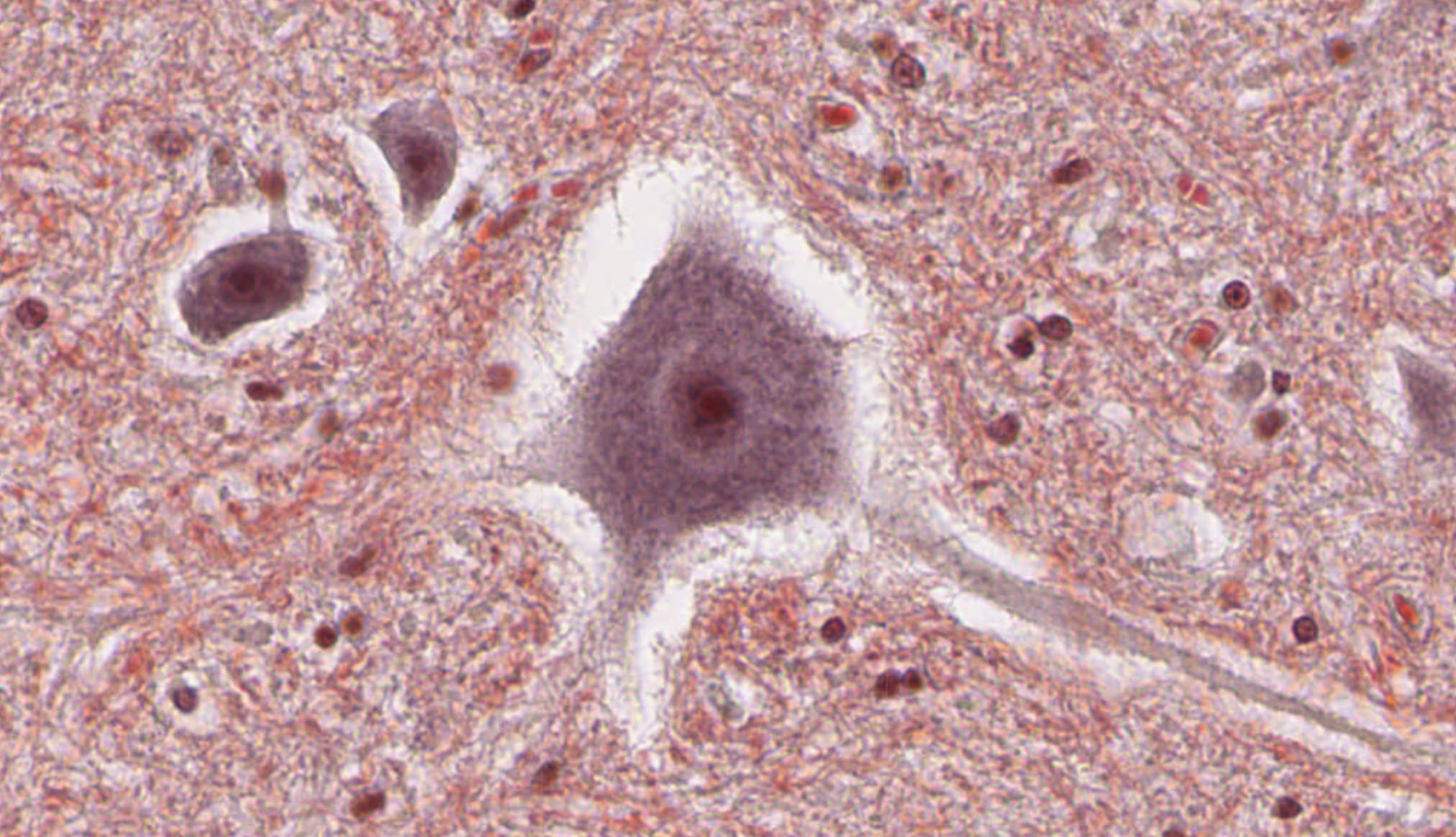
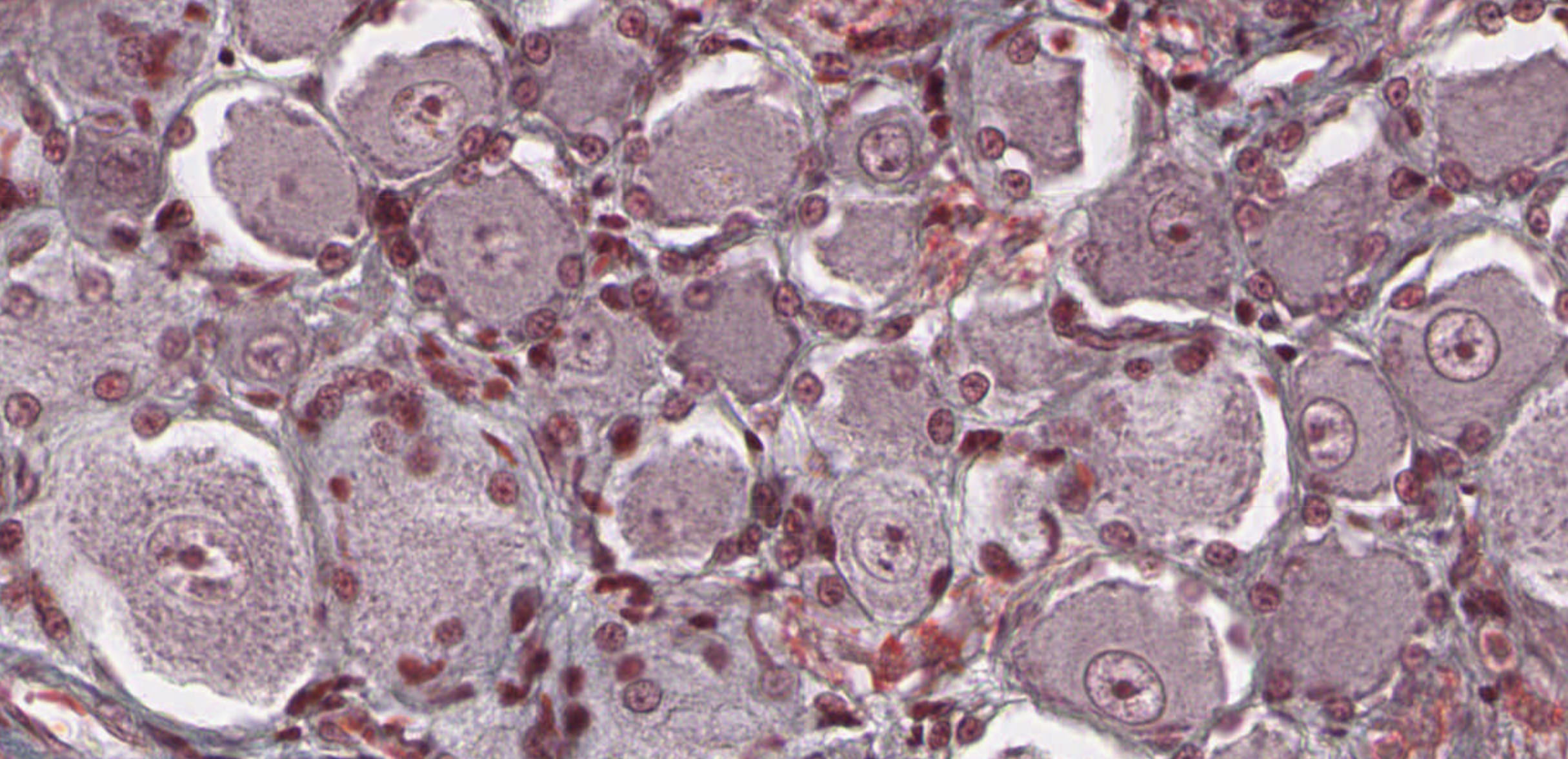
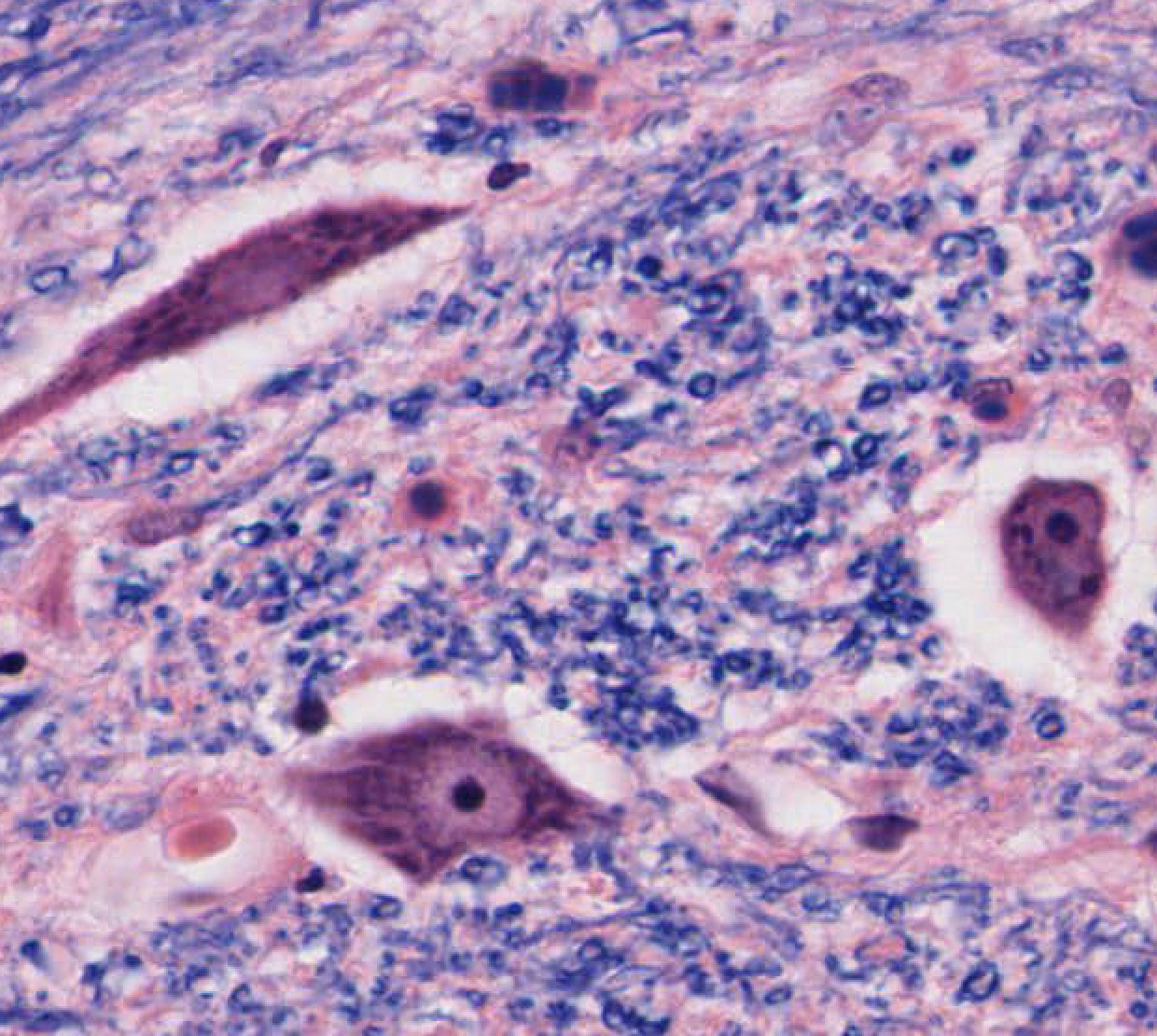
Due to their size and the limited resolution of light microscopy, most cellular organelles are not visible or their detailed structure can't be studies in regular stained tissue sections. The major exception is the cell nucleus of all nucleated cells. This is mainly due to its size and to its content, as nucleic acids are highly basophilic. In larger cells, such as oocytes and many neurons, additional details and substructures of nuclei can be analyzed by light microscopy. Look in the following two slides for Oocytes in the ovary: slide 239 Oocytes View Image, slide 239 Oocytes View Image and for large motor neuron cell bodies in slide 065-1 grey matter of the ventral horn View Image and in slide 065-1 dorsal root ganglia View Image . Find large nuclei and study the distribution of euchromatin and heterochromatin. Note that due to the overall size of these cells, the nucleus may not always be in the plane of sections. In some nuclei you may also be able to detect an intensely stained basophilic nucleolus.
Slide 190B pancreas (rat) chrome-alum hematoxylin and phloxine stain View Virtual Slide
Slide 066a Spinal cord, cross section Masson View Virtual Slide
The detailed structure of the rough endoplasmic reticulum can't be studied by light microscopy. However, the abundance of membrane-bound ribosomes makes areas of rER extremely basophilic and therefore visible, especially in cells that are highly secretory. Good examples are exocrine pancreatic acinar cells and neurons, in which it is referred to as Nissle substance slide 066a Nissle substance View Image. In some histological sections Golgi complexes appear as a pale or slightly eosinophilic (=eosin "loving", an area rich in membranes containing basic amino acids, syn. = acidophilic) regions adjacent to the cell nuclei. In some plasma cells it can sometimes be found in the form of a fine crescent adjacent to the nucleus. However, it takes some practice to recognize it.
Slide 190B pancreas (rat) chrome-alum hematoxylin and phloxine stain View Virtual Slide
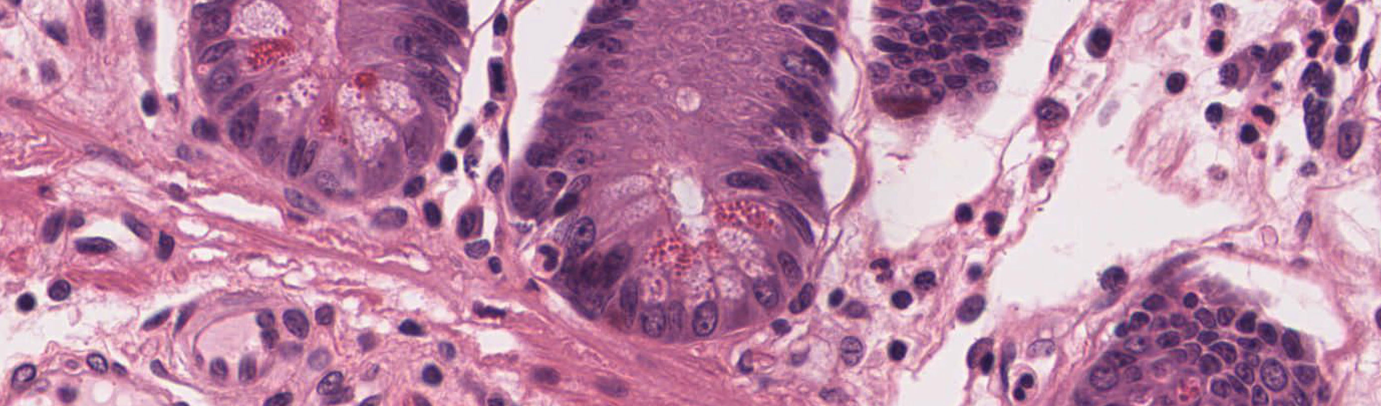
In some secretory cells secretory vesicles can be visualized, especially if their content can be stained with regular histological dyes. View acinar cells in the exocrine pancreas and also the example of some paneth cells in the small intestine slide 029-1 Paneth cells View Image. Note the accumulation of secretory vesicles towards the lumen of the spaces, into which the secretory products will be released.
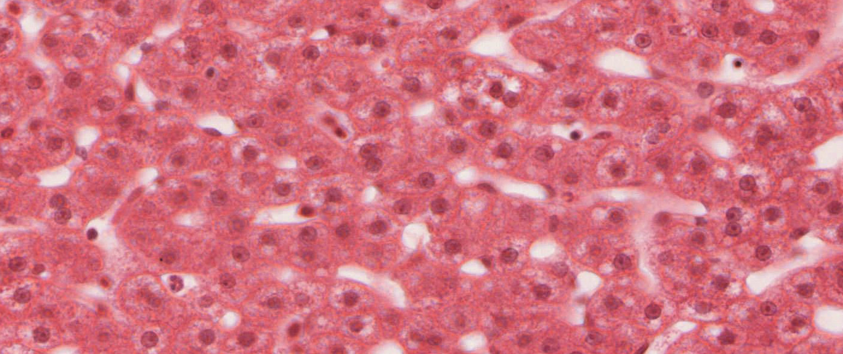
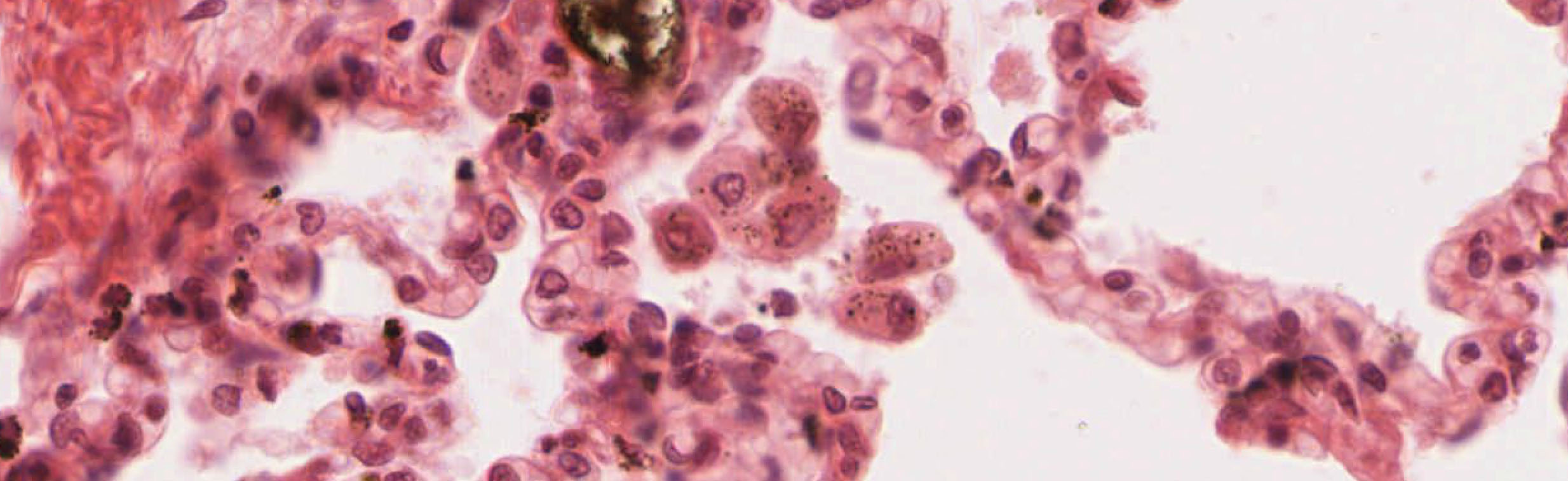
Again, individual endosomes and lysosomes are not visible using regular light microscopy. However, in some cell types, such as macrophages, these cellular compartments show up in regular histological sections as granular inclusions in the cytoplasm. Study the examples of slide 194 macrophage cells in liver View Image macrophage cells in the liver and especially in the lung slide 130-1 macrophage View Image.
Slide 040 trachea H&E View Virtual Slide
Some apical specializations of epithelial cells are visible by light microscopy. Specifically when they are abundant. Due to their size, most cilia are easily recognizable. Identify cilia on the surface of respiratory epithelium. Mobile cilia contain a characteristic set of 9+2 microtubules, which can easily be recognized by electron microscopy and convey an ATP-driven active mobility to these cilia. Most eukaryotic cells also have non-mobile cilia (primary or monocilia), which appear to have a sensory function and have a 9+0 microtubule pattern lacking the central microtubule dimer, e. g. the kinocilium of hair cells. Slide 029-1 Small Intestine (simple columnar epithelium, simple squamous epithelium) H&E View Image. Single microvilli are usually too small to be visible by regular light microscopy. However, they become visible as a collective when they forming a dense layer at the apical surface of mostly absorptive epithelia (e. g. kidney tubules and intestine). These structures are referred to as brush or striated border. A second type of actin-containing apical protrusion are stereocilia. In size they approach the dimension of cilia and are readily visible by regular light microscopy. They can be found on sensory hair cells and the epithelium of the epididymis (Look at the orientation slide to find the region of the epididymus).
Slide 112 thick skin, sole of foot View Virtual Slide
Most cell junctions are not visible using light microscopy. The spines in the stratum spinosum of the skin are one notable exception. These spines represent desmosomes that hold skin epithelial cells together.
21 Plasma cell View Virtual EM Slide
This electron micrograph shows a typical secretory cell, a plasma cell, which secretes immunoglobulin protein. Many of the major types of cellular organelles are visible in this image. In the nucleus, areas of euchromatin and heterochromatin can easily be identified. Use these micrographs to review the structure of organelles. Be sure you recognise favourable sections of the nucleas, mitochondria, and rough ER.
11 Nuclear Envelope - Transverse Section View Virtual EM Slide
This electron micrograph nicely depicts the double membrane of the nuclear envelope including some nuclear pores.
12 Pancreas Rough ER View Virtual EM Slide
This electron micrograph shows the typical flat cisternae of the rough endoplasmic reticulum, which are studded with ribosomes.
13 Golgi apparatus - Exocrine Pancreas View Virtual EM Slide
The Golgi apparatus looks rather unusual in this electron micrograph. This is due to the enlarged stacks of cisternae (Golgi vacuoles), which distort the appearance of the Golgi complex.
227 Pancreas - Exocrine, detail of acinus Organelles of the Secretory Pathway &View Virtual EM Slide
Pancreatic acinar cells as depicted in this electron micrograph are cells that are highly specialized for protein secretion. Therefore, all the organelles of the protein secretory pathway are well-represented and are clearly visible in this micrograph.
14 Centrioles View Virtual EM Slide
This image shows two centrioles, which represent the central structure of the microtubule-organizing center (MTOC). Some microtubules are also visible in the vicinity.
155 Cilia View Virtual EM Slide
Cross sections of cilia. The typical 9+2 arrangement of the microtubules is especially evident in EM #156.
156 Cilia - Cross-sectioned in human trachea View Virtual EM Slide
Cross sections of cilia. The typical 9+2 arrangement of the microtubules is especially evident. The basal bodies are centrioles and have 9 triplets of microtubules with no central pair.
170 Kidney - Cortex, proximal tuble Brush Border View Virtual EM Slide
This EM micrograph depicts the typical appearance of microvilli on the apical surface of two types of cells with a striated or brush border. Shown is the epithelial lining cell of a proximal tubule in the kidney.
213 Small intestine - microvilli View Virtual EM Slide
Study the typical appearance of microvilli on the apical surface of this cell forming part of the striated or brush border in small intestine.
16 Epithelium - Desmosome and Intermediate Filaments View Virtual EM Slide
A desmosome can be seen in the upper right corner of this transmission electron micrograph. The cytoplasm is full of intermediate filaments (tonofilaments), some of which are attached to the desmosomal plaque.
Click on a question to reveal the answer.
In which cell organelle(s) occur(s) the addition and modification of carbohydrate moieties of secretory and membrane proteins
N-linked glycosylation usually happens co-translational at the level of the rough endoplasmic reticulum. O-linked glycosylation and modifications of N-linked carbohydrate moieties are functions of the Golgi complex.
Describe the two major pathways of protein degradation in cells.
Extracellular and membrane proteins are taken up by cells using a vesicle- mediated process (usually coated vesicles) and are degraded via the endosome and lysosomal pathway. Many intracellular proteins, which are destined for degradation, are first covalently modified with multiple ubiquitin peptides. Subsequently they are recognized and proteolytically degraded by cytosolic proteasomes.
1. Which of the following cell organelles is NOT involved in the synthesis, processing and export of a secretory protein?
- Golgi complex
- Trans-Golgi network
- Endosome
- Secretory granules
- Plasma membrane
- Rough endoplasmic reticulum
Answer
Correct answer 3. Endosomes are part of the endocytosis pathway, not the secretary pathway.
2. Which of these microscopy methods has the highest resolution (is able to depict the smallest structures)?
- Differential interference light microscopy
- Bright field light microscopy
- Fluorescence microscopy
- Transmission electron microscopy
- A magnifying glass
- Phase contrast microscopy
Answer
Correct answer 4. Transmission electron microscopy as it uses very short wave electrons, rather than longer wave electromagnetic radiation (visible light or UV)
3. Which type of protein is usually NOT synthesized by free ribosomes in the cytoplasm?
- Nuclear proteins
- Secreted proteins
- Cytoplasmic proteins
- Most Mitochondrial proteins
- Most Chloroplast proteins
- Peroxisomal proteins
Answer
Correct answer 2. Secreted proteins are synthesized by rough ER membrane-bound ribosomes.
4. Which of the following structures or terms is NOT associated with the proteolytic destruction of cellular proteins?
- Ribosomes
- Lysosomes
- Endosomes
- Ubiquitin
- Proteasomes
Answer
Correct answer 1. Ribosomes are involved in synthesizing proteins, not to degrade them.
5. Which of the following statements about mitochondria is FALSE?
- Mitochondria have two membranes/lipid bilayers
- The inner mitochondrial lipid bilayer contains a complex of electron-transporting proteins
- Mitochondria contain ribosomes, which synthesize mitochondrial proteins
- A proton gradient across the inner mitochondrial lipid bilayer drives the generation of ATP
- Mitochondria may be derived from fungal eukaryotic cells that have been taken up by a eukaryotic cell
Answer
Correct answer 5. Mitochondria are NOT derived from fungal eukarotic cells that have been taken up by a eukaryotic cell. Fungi are deukaryotic cells that have their own mitochondria. The endosymbiotic hypothesis states that cyanobacteria were taken up by early eukaryotic cells and over time converted into mitochondria and chloroplasts.
6. Which of the following terms does NOT describe a cytoskeletal structure/protein complex?
- Nucleosomes (Histones)
- Microfilaments (Actin)
- Membrane skeleton (Spectrin-ankyrin)
- Microtubules (Tubulin)
- Intermediate filaments (Keratin, Neurofilaments etc. )
Answer
Correct answer 1. Nucleosomes (Histones) are responsible for DNA packaging in the nucleus.