- Be able to identify cells and tissues in the peripheral nervous system (nerves, neurons and glia).
- Describe the organization of a typical neuron and the direction of information flow.
- Describe and contrast the function and organization of sensory and motor neurons.
- Describe the process of myelination, and the function of myelin, including Nodes of Ranvier. Explain the role of the Schwann cell, with respect to both myelinated and unmyelinated neurons.
- Describe the organization of connective tissue in a nerve.
- Know the function and basic organization of neuromuscular junctions (motor end plates) and muscle spindles.
The nervous system is divided structurally into the central nervous system (CNS) and the peripheral nervous system (PNS), although remember that these are really two components of one, integrated system. The CNS consists of the brain and the spinal cord, while the PNS is composed of nerves and groups of nerve cells (neurons), called ganglia. The nerves of the PNS carry sensory (afferent) inputs to the CNS and motor (efferent) output from the CNS to the skeletal and cardiac muscles and the smooth muscles of blood vessels, organs and glands. You will learn more about the histological organization of the CNS in detail later in the year. However, in this lesson you will study the spinal cord briefly because the cell bodies (perikarya) of motor (efferent) nerve fibers in the peripheral nerves are located in the spinal cord. Another way to think about the nervous system is according to its two main functional subdivisions: the somatic (voluntary, conscious) and autonomic (involuntary, unconscious) components. For additional information please review the "Overview of the Nervous System" module from the Gross Anatomy website.
Slide 65-1N (spinal cord, trichrome) View Virtual Slide
Slide 65-2 (spinal cord, H&E) View Virtual Slide
Slide 066a thoracic spinal cord thoracic spinal cord luxol blue cross View Virtual Slide
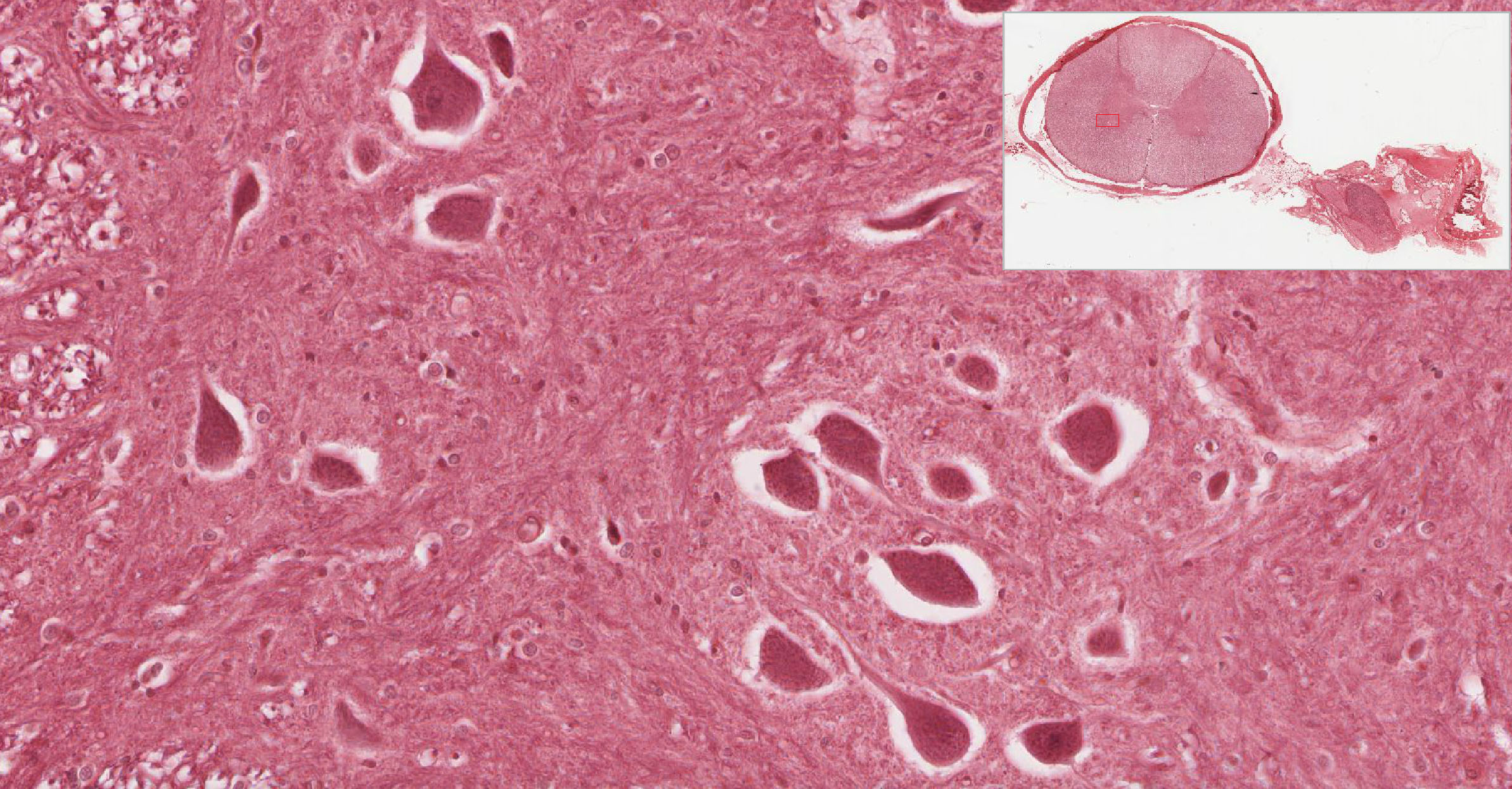
Review the organization of the spinal cord using your atlas. Examine the cross section of the spinal cord in slide #65-2. At low magnification, differentiate inner gray from outer white matter and identify dorsal and ventral horns of the gray matter. You should also identify the dorsal and ventral horns in slide 65-1N stained with Masson trichrome. In these slides, dorsal happens to be "up," but you should be able to tell dorsal and ventral horns based on morphology and the cells present rather than the orientation. The perikarya of large somatic motor neurons View Image located in the ventral horn of the cord innervate the skeletal muscles of the limbs and trunk, which are embryologically derived from somites (hence, "somatic" muscles). Observe that the perikarya of neurons in the dorsal horn are much smaller. Why are the neurons of the dorsal horn smaller?
Answer
Neurons in the dorsal horn are essentially interneurons that project to other regions of the CNS (e.g. motor neurons in the spinal cord or sensory input to the brain), so they have much smaller overall volume and therefore much less metabolic demand compared to motor neurons which project to target muscles that may be more than a meter away. Remember that the perikaryon is the metabolic support center for each neuron, so, therefore, motor neurons require much larger perikarya
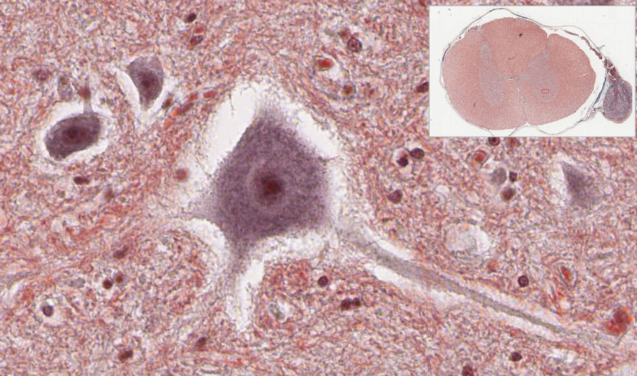
Many neurons in the spinal cord may appear shrunken and surrounded by an empty space due to poor fixation. Cells that are well preserved show features characteristic of most neurons: large cell body, large pale nucleus, Nissl substance, and cell processes (most of which are dendrites). The delicate meshwork of dendritic processes and nervefibers (axons) lying between cells in the gray matter is called the neuropil. The white matter contains nerve fibers (axons) entering and exiting the gray matter, and traveling up and down the spinal cord, linking it to the brain. Nervous tissue contains two basic categories of cells: neurons and support cells (glia). Both neurons and glia have fine processes projecting from the cell body, which generally cannot be resolved in the light microscope without special staining techniques. In the CNS,oligodendrocytes (a type of glial cell) are responsible for myelination of CNS axons. We will consider the process of myelination below, in the discussion of Schwann cells, which are glial cells that produce myelin in peripheral nerves. Myelin is lipid-rich, and on gross inspection appears white. Thus, in the 'white matter' of the brain and spinal cord, myelinated axons are the predominant neuronal component, whereas ‘gray matter’ contains relatively more neuronal and glial perikarya and non-myelinated (e.g. dendritic) processes.
Neurons are characterized by a large cell body or perikaryon containing a large, pale (active, euchromatic) nucleus with a prominentnucleolus. Scattered in the cytoplasm are the characteristic clusters of ribosomes and rough ER termed Nissl bodies or substance View Image. One or more cell processes may also be seen emerging from the neuronal perikaryon. Review diagrams illustrating the morphology of neurons in your textbooks. The dendrites receive neural input from other neurons via synapses (or they are specialized to receive sensory stimuli), and they transmit neural information toward the perikaryon. A single axon (often called a nerve fiber) leaves the perikaryon and transmits neural signals to other neurons or to the effector organ (e.g., skeletal muscles) via synapses. The specialized synaptic connection between axons and their target muscle fibers is called a neuromuscular junction, which will be discussed below.
In the peripheral nervous system, clusters of neurons with associated nerve fibers and supporting cells are referred to as ganglia. (In the central nervous system, clusters of neurons are referred to as "nuclei", an unfortunate terminology.)
A.
Dorsal Root (Spinal) Ganglia Slide 65-1N (spinal cord, trichrome) View Virtual Slide
Slide 65-2 (spinal cord, H&E) View Virtual Slide
Slide 65-1 (spinal cord, trichrome) View Virtual Slide
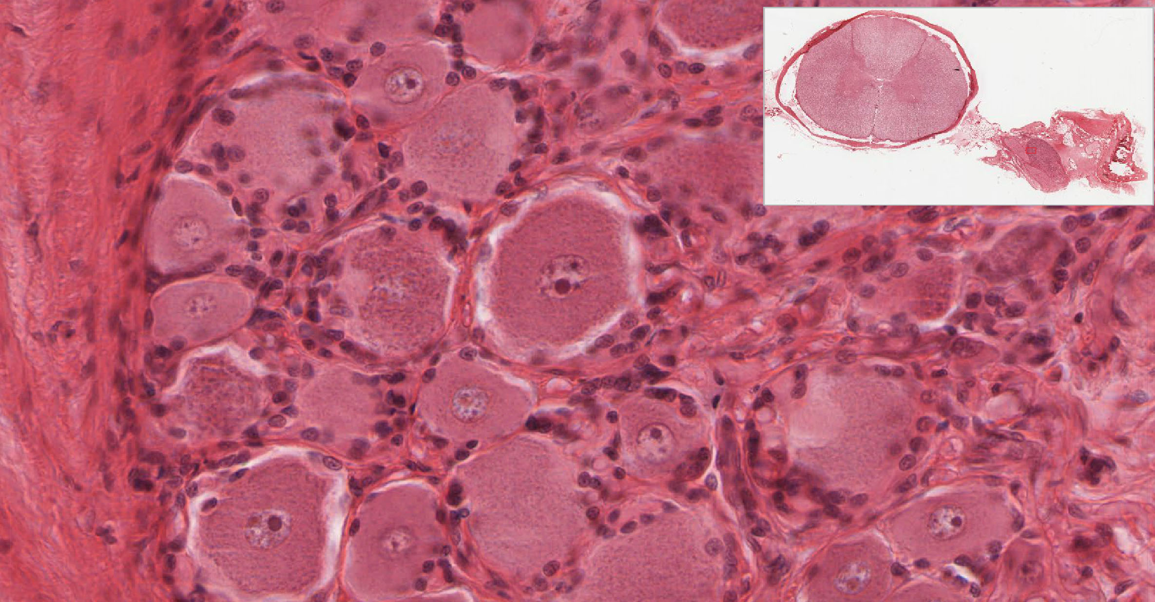
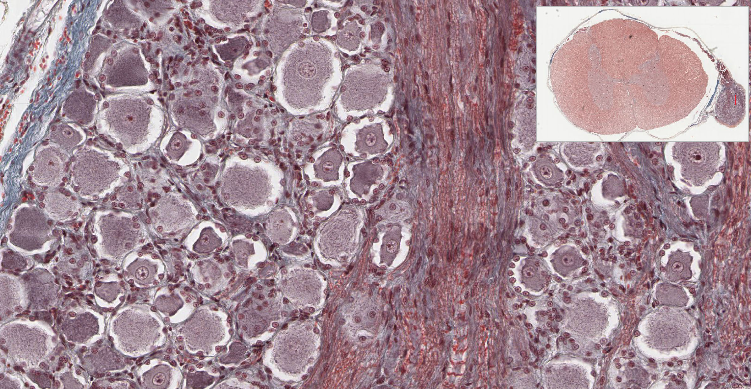
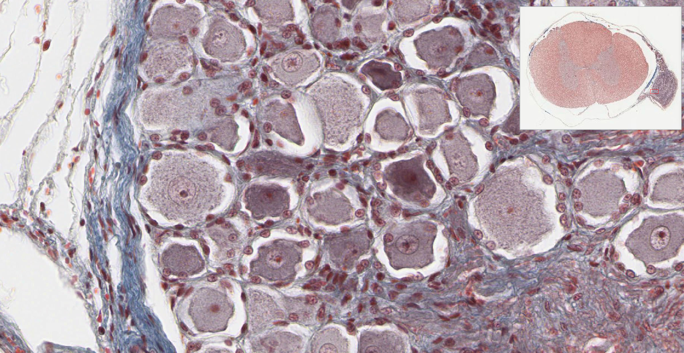
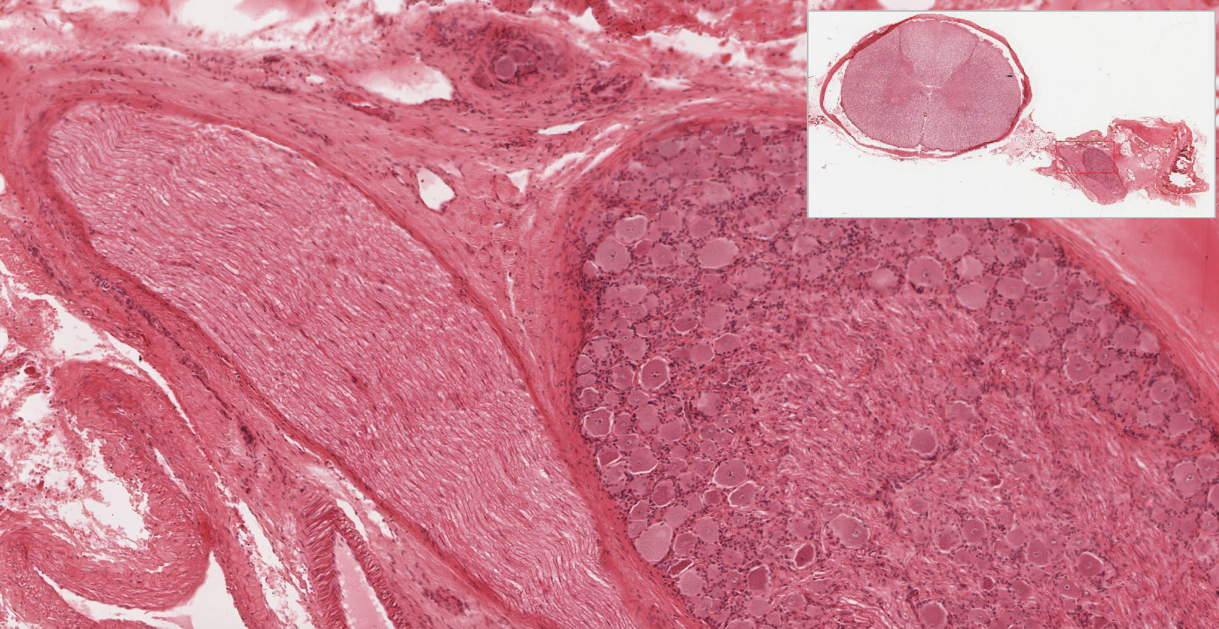
Dorsal root ganglia contain the cell bodies of sensory neurons. Return to slide #65 and locate a dorsal root ganglion near, but outside, the spinal cord. The neuron cell bodies belong to large, pseudounipolar sensory neurons that have a single "T-shaped" process; these are theafferent fibers carrying sensory information from the periphery (sensory receptors in the skin, joints and muscles that respond to touch, temperature, pain, stretch) to the dorsal horn, where they synapse on neurons in the spinal cord. NOTE: these sensory neurons are an exception to the typical neuron, in that they do not have separate dendrites and an axonal process, but rather one branched process that serves both functions. Many of the clusters of sensory neuron cell bodies are peripheral in the ganglion, and others lie between bundles of nerve fibers running in parallel through the ganglion. There are no synapses in these ganglia. You will seldom see a process coming from the cell body, since cells are pseudounipolar and the process will not usually be included in the plane of section. The nuclei in dorsal root ganglia are generally located centrally in the cell bodies of the neurons. Numerous satellite cells (a type of glial cell) form a prominent capsule around each cell body evident in H&E-stained slide 65-2 View Image and Masson-stained slides 65-1 View Image and 65-1N View Image. Just as in the spinal cord, many neurons may appear shrunken and surrounded by an empty space due to poor fixation. The perikarya of surrounding glial cells are typically much smaller than neurons, and their nuclei contain markedly less euchromatin.
B.
Autonomic ganglia Slide 74 (sympathetic ganglion, toluoidine blue) View Virtual Slide
Slide 250-1 (vagina, H&E) View Virtual Slide
Slide 250-2 (vagina, trichrome) View Virtual Slide
Slide 75 (seminal vesicle, H&E) View Virtual Slide
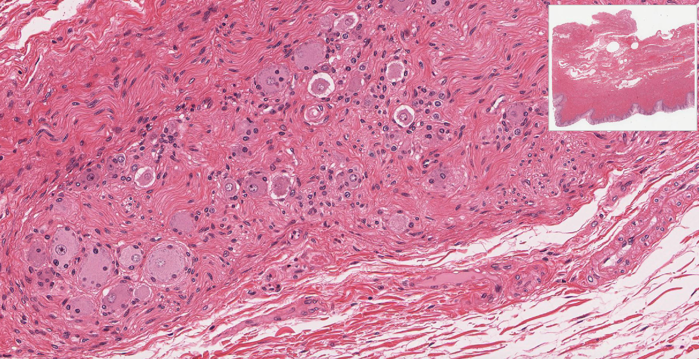
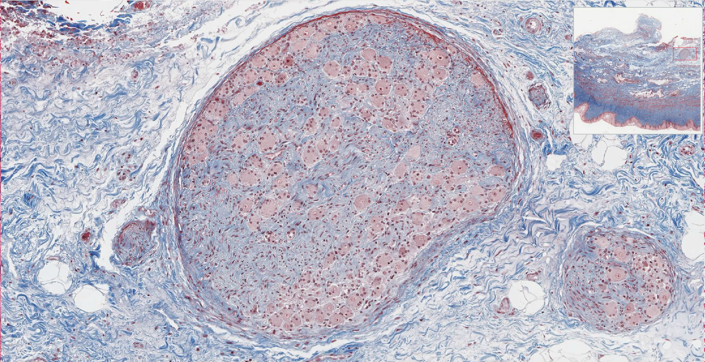
Autonomic ganglia contain cell bodies of sympathetic or parasympathetic motor neurons, which receive synaptic input from preganglionic autonomic neurons whose cell bodies are located in the CNS. The autonomic motor neurons in the ganglia send efferent fibers (postganglionic autonomic nerve fibers) to innervate cardiac muscle fibers of the heart and smooth muscle fibers of body organs and glands. Examine a sympathetic ganglion in slide #74. Compare this autonomic ganglion with the dorsal root (spinal) ganglia studied above. The neuron cell bodies are often more widely dispersed, with a meshwork of nerve fibers lying between them, and the nerve fibers generally are not as well organized. Unlike the dorsal root ganglia, which have no synapses and therefore no neuropil, in sympathetic ganglia many preganglionic sympathetic fibers from the spinal cord synapse on the sympathetic neurons, and others travel through the ganglia without synapsing. The cell bodies of sympathetic neurons are smaller than those of sensory neurons in the dorsal root ganglion, and often have eccentrically placed nuclei. The cell profile appears somewhat angular, since these cells are multipolar, and the roots of their processes are often included in the plane of section. The satellite cells (glial cells) are sparse and less apparent. Parasympathetic ganglia are located in the organ that is being innervated. Go to slide #250 (vagina) stained with H&E View Image or Masson trichrome View Image or slide #75 (seminal vesicle) View Image and see if you can identify parasympathetic ganglia amongst large blood vessels and nerves in the deep connective tissue in the vaginal wall (outer 1/3), or in the connective surrounding the seminal vesicle.You should be able to distinguish dorsal root (spinal) ganglia from autonomic ganglia and to identify neurons and satellite cells in these ganglia. What neuronal perikarya are found outside the CNS? NS2).
Answer
Most neuron cell bodies (perikarya) are within the CNS (central nervous system, i.e. brain and spinal cord). This includes the perikarya of motor (efferent) neurons located in the ventral (anterior) horn of the spinal cord, but whose axons exit in the spinal nerves to innervate voluntary (skeletal) muscles. The cell bodies of sensory (afferent) and autonomic (visceral motor) neurons lie outside the spinal cord, in small aggregates of neurons and support cells called ganglia. The perikarya of sensory neurons are in the dorsal root ganglia. These pseudounipolar neurons have a T-shaped process, which carries sensory information from the periphery to the spinal cord. Perikarya of the autonomic (sympathetic and parasympathetic) nervous system neurons are found in ganglia near the spinal cord (sympathetic) or in the target organs (parasympathetic).
Slide 68 (myelinated nerve, trans. section, H&E) View Virtual Slide
Slide 67 (myelinated nerve, long. section, H&E) View Virtual Slide
Slide 65-2 (spinal cord, H&E) View Virtual Slide
Slide 29 (intestine, trans. section, H&E) View Virtual Slide
Slide 155 (gastro-esophageal junction, long. sect., H&E) View Virtual Slide
Slide 169 (jejunum, trans. section, H&E) View Virtual Slide
In the peripheral nervous system, the larger diameter axons are surrounded by a lipid-rich myelin sheath formed by the Schwann cells(Wheater's pg. 138, 7.18). The Schwann cells (in the peripheral nerves) and the satellite cells (in the ganglia) are glial cells (supporting cells) of the PNS. Using slide #68, examine a cross section of a nerve trunk. It is made up of several fascicles, two of which are larger than the others. Within one of the larger fascicles, study the axons in cross section, noting the wide range in axon diameter. The axon appears as a dot in the middle of a clear space, which is the region occupied by lipid-rich myelin (extracted during tissue preparation). Most peripheral nerves carry both afferent (sensory) and efferent (motor) fibers. The nerve and the fascicles (bundles of nerve fibers) that comprise it in this section are invested with a thick layer of dense connective tissue or epineurium. Each fascicle is surrounded intimately by the perineurium, which is a layer of dark-staining, flattened cells lying between the epineurium and groups of axons of the fascicle. The endoneurium is a delicate layer of reticular fibers and other connective tissue components surrounding each individual axon. What distinguishes endoneurium, perineurium and epineurium?
Answer
Nerves that are visible upon gross dissection are covered with epineurium, a layer of loose collagenous tissue that bundles several fascicles, each containing many nerve fibers. The individual nerve fascicles are contained within a condensed layer of collagenous tissue called the perineurium. Blood vessels run longitudinally within compartments formed by epineurium and perineurium. Within the perineurium, nerve fibers and their ensheathing Schwann cells are surrounded by endoneurium, a delicate layer of connective tissue with a capillary network, separated from the Schwann cell by a basement membrane.
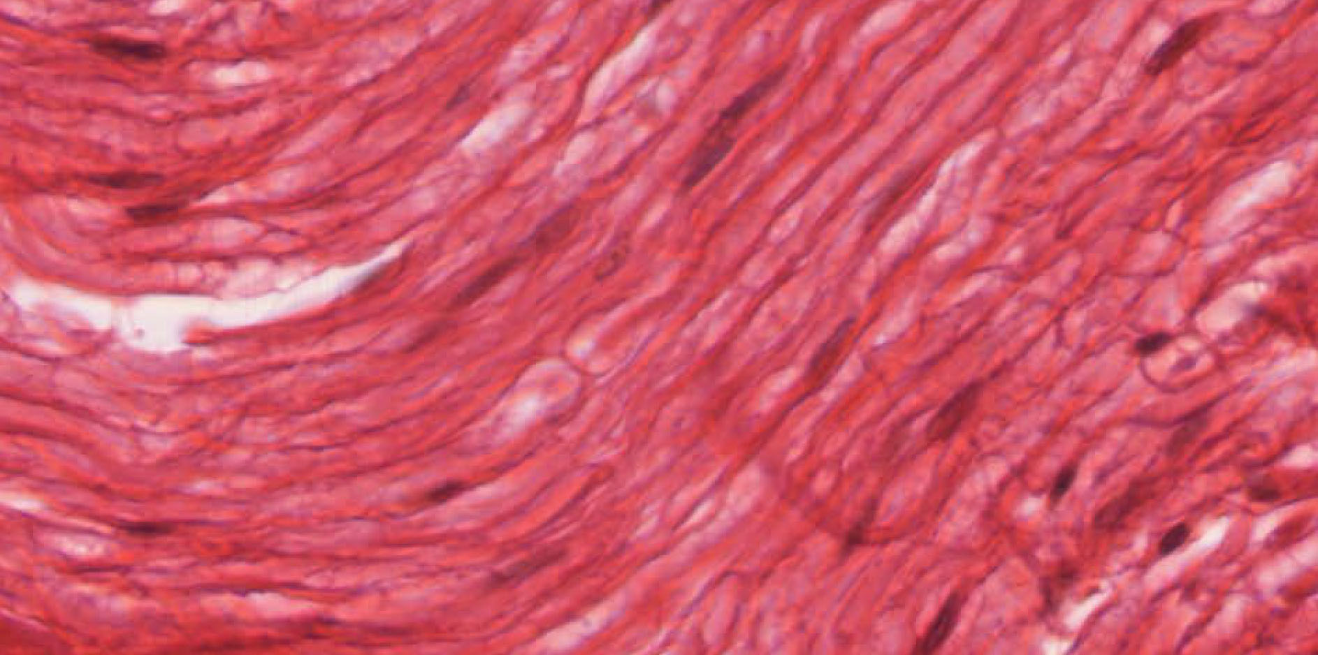
Next, examine a longitudinal section of peripheral nerve, slide #67. Note the wavy course of the myelinated axons, which is characteristic of nerves seen in histological sections. Note that you can see the slightly darker axon running through the clear myelin space. Look for nodes of Ranvier View Image in places where the axons are cut in particularly favorable longitudinal section. Myelinated axons may also be found in the dorsal nerve root in slide 65-2 View Image. As before, the nuclei present among the axons belong mostly to Schwann cells or to fibroblasts, although there are also occasional capillaries. What are the numerous nuclei observed in non-myelinated nerves?
Answer
Though instinctively you may guess that these are nerve cell nuclei, the nuclei of nerve cells are generally located in ganglia or in the spinal cord, not randomly throughout the nerve. Also, nerve cell bodies are huge and very obvious. Instead, the nuclei observed in non-myelinated nerves are mostly Schwann cells (there are occasional fibroblasts as well). There are two ways that Schwann cells interact with nerves. They can myelinate the nerve or they can just envelop the nerve without myelinating it. Many non-myelinated nerve fibers can be enveloped in a Schwann cell, and non-myelinated fibers tend to be smaller in diameter than myelinated fibers
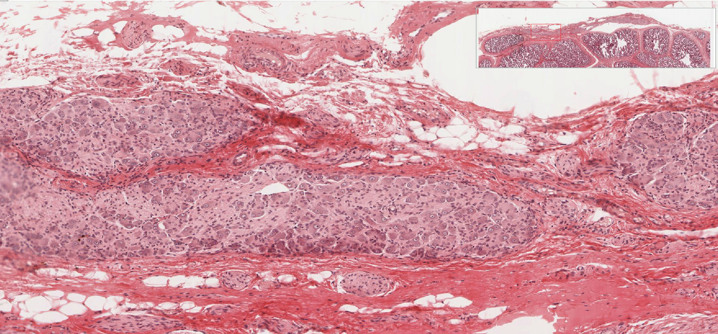
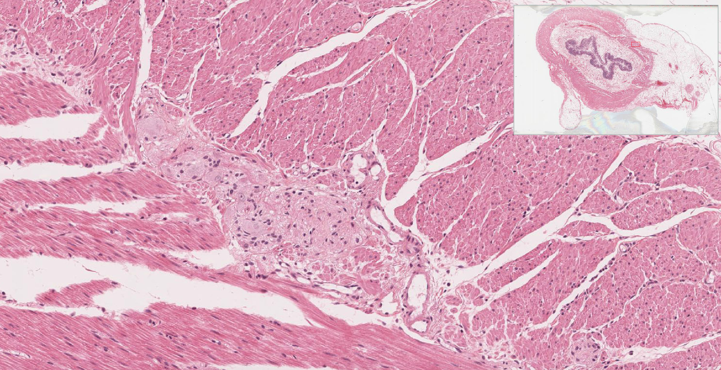
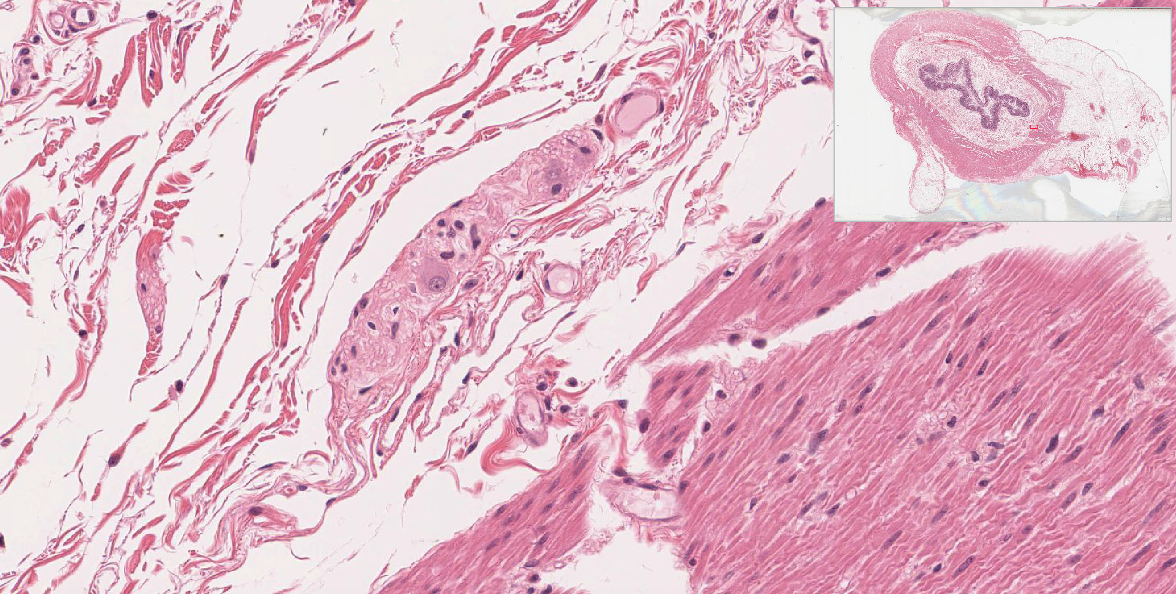
Examine slides #29, #155, and #169 to find small nerve fibers and more autonomic (parasympathetic) ganglia. Look in between the layers of smooth muscle you studied in the connective tissue lab session. An extensive plexus of nerves and parasympathetic ganglia (myenteric plexus) is present in the connective tissue separating these muscle layers, shown particularly well in slide 29 View Image and slide 155. Identify both nerve fibers and neurons of the parasympathetic ganglia. This is a good way to practice distinguishing smooth muscle and connective tissue from adjacent nerve fibers. Small parasympathetic ganglia and nerve fibers may also be found in the connective tissue of the submucosa in slide 29 View Image and slide 169.
Slide 71-2A (motor end plates, Golgi colloidal gold stain) View Virtual Slide
Slide 71-1B (muscle and muscle spindle, trans. section, H&E) View Virtual Slide
Slide 71-1A (muscle and muscle spindle, trans. section, H&E) View Virtual Slide
Lower/Alpha (somatic) motor neurons from the ventral horn of the spinal cord innervate muscle fibers (their effector cells) at specialized synapses called neuromuscular junctions (or motor end plates of skeletal muscles) View Image. These are best visualized with special stains that use heavy metals (gold) to label the nerve fibers or histochemical methods for acetylcholinesterase (an enzyme that hydrolyzes the neurotransmitter used by somatic motor neurons, acetylcholine). The slide #71 in even numbered boxes (digital slide 71-2A) show a similar preparation of motor end plates. Also, review the diagrams in your atlas. The terminal bouton of the motor axon has numerous synaptic vesicles that contain the neurotransmitter, acetylcholine. The terminal bouton lies in a depression in the surface of the muscle fiber, and is separated from it by a gap, the synaptic cleft, of uniform width. The plasma membrane of the muscle fiber is highly folded, and a basal lamina (also called the external lamina), is interposed between the nerve fiber and muscle fiber. Synaptic transmission of nerve impulses across the synaptic cleft is accomplished by the release of acetylcholine from the synaptic vesicles (by exocytosis) into the synaptic cleft, where it diffuses to the muscle fiber membrane and activates acetylcholine receptors, which trigger membrane depolarization and subsequent muscle contraction. Neuromuscular spindles are stretch receptor organs that regulate muscle tone via the spinal stretch reflex. Look at slide #71 in odd-numbered boxes (digital slide 71-1B) and identify the neuromuscular spindle View Image in the belly of the muscle (look within the perimysium between the muscle fascicles [see orientation]). In this preparation (H&E, transverse section), the sensory nerve fibers of the spindle are not visible, but the modified skeletal muscle fibers (intrafusal fibers), which are smaller than the muscle fibers proper (extrafusal fibers), are easily visualized - 2 to 10 are contained in a fluid-filled space within a discrete, external connective tissue capsule. Note the intrafusal fibers are bundled together by a delicate internal capsule that is not so evident in these sections. The sensory receptors (nerve endings) are activated by stretching of the intrafusal fibers, which evokes a reflex contraction of the extrafusal fibers that is driven by large (alpha) somatic motor neurons (located in the ventral horn) in a two-neuron spinal reflex arc. It is worth noting that, in addition to being stretch receptors, the intrafusal fibers are functional, contractile muscle cells. They are innervated by special (gamma) motor neurons that set the tone of the intrafusal fibers thus modulating sensitivity of the stretch receptor (contraction of the spindle cells makes them more taut and therefore even more sensitive to stretch). This also allows the spindle cells to contract in concert with the extrafusal fibers thus maintaining sensitivity to stretch over the muscle's full range of motion [see explanatory figure].
![[see orientation]](/sites/default/files/images/slides/15PNS.jpg)
![[see explanatory figure]](/sites/default/files/images/slides/16PNS.jpg)
49 Motor nerve cell - Ventral Horn of Rabbit Spinal Cord, Multipolar Motor Neuron Cell Body View Virtual EM Slide
Motor Neuron Cell Body. In this electron micrograph, note some of the features you saw in ventral horn motor neurons with the light microscope, such as the large, pale nucleus, prominent nucleolus, Nissl bodies, dendrites and axon. Adjacent to the neuron, note myelinated axons of various sizes and also that there are no spaces between cell processes. All spaces are occupied either by the processes of neurons or glia or by capillaries (these capillaries are somewhat swollen here because the tissue was fixed by perfusion).
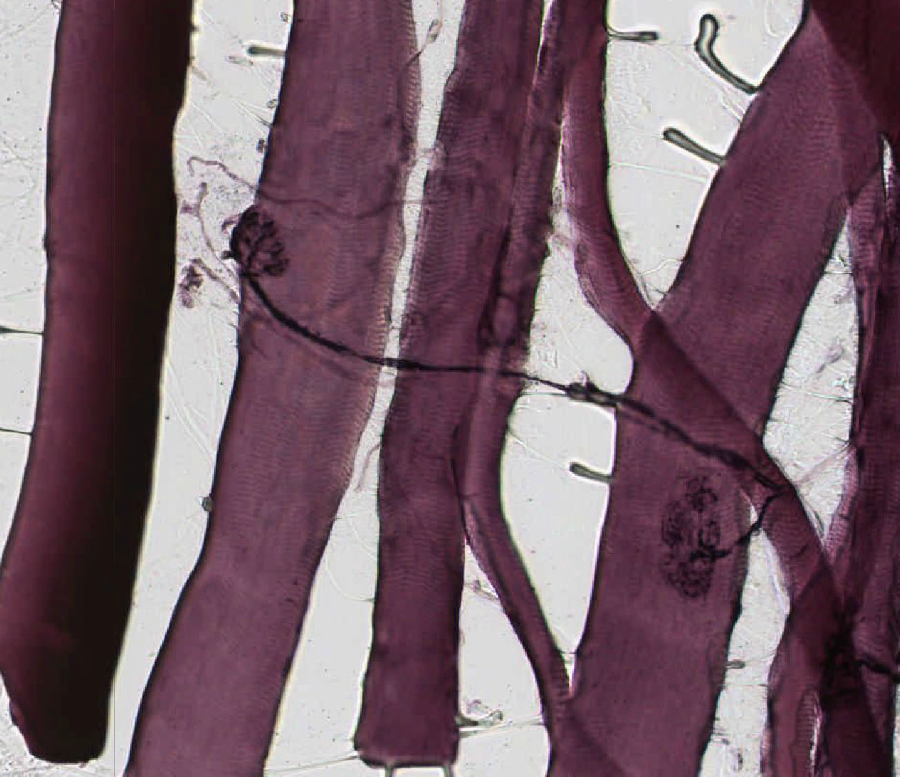
52 Peripheral nerve - Longitudinal section of Node of Ranvier View Virtual EM Slide
Node of Ranvier (longitudinal section). Remember that the node of Ranvier is actually a short segment of the axon that is bare at the junction between two Schwann cells, making "saltatory conduction" possible. Note the manner in which the myelin ends in each Schwann cell at the junction, by a "peeling off" of successive myelin layers, which come to lie against the axon as small cytoplasmic swellings.
53 Peripheral Nerve - Longitudinal section - Myelinated View Virtual EM Slide
Myelinated Nerve Fibers (longitudinal section). This image shows the typical appearance of a myelinated nerve, consisting of parallel bundles of axons (light areas) wrapped with sheaths of myelin (dark areas). As in the peripheral nervous system each Schwann cell myelinates only one axon, the discontinuous appearance of the axon labeled 5 in a Schwann cell is probably due to its curvature around the nucleus.
54 Peripheral nerve -Schwann Cell with Myelinated Nerve Fiber View Virtual EM Slide
Schwann Cell with Myelinated Nerve Fiber (cross section). In this cross section of a myelinated nerve process, note the axon, containing microtubules and neurofilaments and bounded by a plasma membrane ("axolemma"). Outside the plasma membrane of the axon is the myelin sheath, which you will remember is composed of tightly wrapped plasma membranes of the Schwann cell. Also, note the nucleus and cytoplasmic organelles of the Schwann cell. Remember that the myelin is part of the Schwann cell, not of the axon.
55 Peripheral nerve - Unmyelinated Nerve Fibers - Cross Section View Virtual EM Slide
Unmyelinated Nerve Fibers (cross section). The axons seen in this electron micrograph are all non-myelinated. They are embedded in grooves in the Schwann cell surface (in some cases there may be more than one axon per groove), with each Schwann cell thus supporting a considerable number of these small, unmyelinated axons. Although the axons are very close together, you will observe thin partitions of Schwann cell between them.
57 Celiac Ganglion - Rabbit - Autonomic - Multipolar Cells Multipolar Neurons View Virtual EM Slide
Multipolar Neurons (Celiac Ganglion). The celiac ganglia are autonomic ganglia. Note the large ganglion cells with somewhat eccentrically placed nuclei in several cells, a characteristic feature of autonomic ganglion cells.
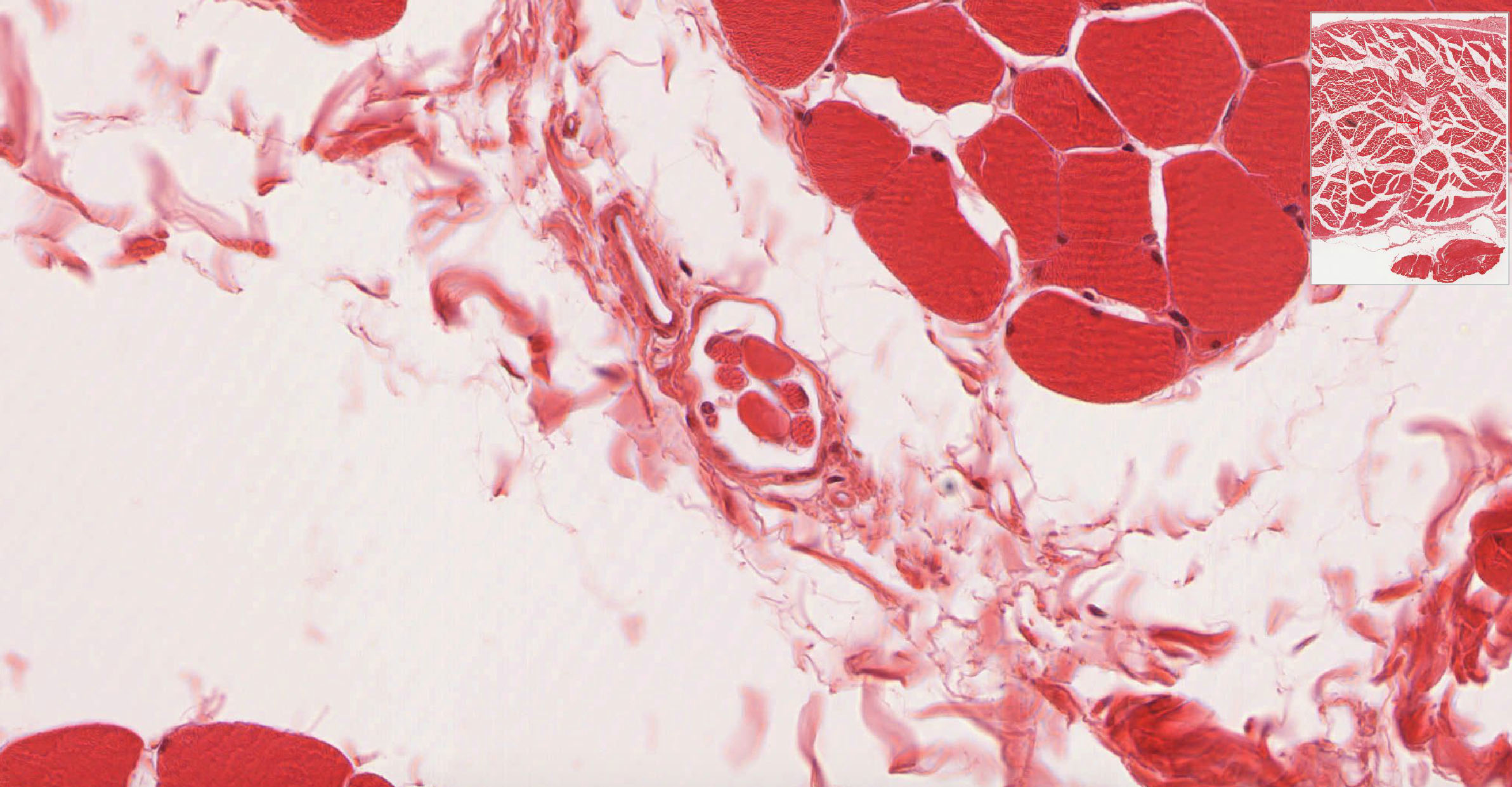
59 Nerve vessel bundle - Cross Section of Rat Skeletal Muscle Myelinated Nerve with Endoneurium and Perineurium View Virtual EM Slide
Myelinated Nerve with Endoneurium and Perineurium (cross section). Between the axons you will see delicate connective tissue and an occasional fibroblast, which constitute the endoneurium. At the periphery of the fascicle, observe the perineurium, made up of several layers of flattened cells; it is a highly specialized layer that acts as a barrier and protects the nerve from the environment.
60 Myoneural junction - Rat Motor End Plate Neuromuscular Junction View Virtual EM Slide
Neuromuscular Junction. This is a motor end plate. Note that the nerve axon loses its myelin sheath as it approaches the motor end plate and it terminates as a bulbous expansion in a trough of the muscle cell surface. The bulbous knob reveals numerous mitochondria and small synaptic vesicles, which contain cholinergic neurotransmitter substances. When these vesicles fuse the cell membrane of the axon bulb (the presynaptic membrane) and the content is released into the synaptic clefts to be taken up by the postsynaptic membrane (muscle cell membrane).
61 Neuromuscular spindle - Cross Section of a polar segment Neuromuscular Spindle View Virtual EM Slide
Neuromuscular Spindle. Note the two types of intrafusial muscle fibers, the nuclear bag fibers and nuclear chain fibers are enclosed by a delicate internal capsule. This arrangement serves as a muscle stretch receptor. The sensory nerve endings are activated by the stretching of the intrafusial fibers and the nerve impulse generated excites the somatic motor neurons in the spinal cord to evoke a reflex contraction of extrafusial fibers.
Click on a question to reveal the answer.
Why are perikarya of dorsal horn neurons smaller than those in the ventral horn?
Neurons in the dorsal horn are essentially interneurons that project to other regions of the CNS (e.g. motor neurons in the spinal cord or sensory input to the brain). As they project to closer targets, they have much smaller overall volume and much less metabolic demand compared to motor neurons which project to target muscles that may be more than a meter away. Remember that the perikaryon is the metabolic support center for each neuron, so, therefore, motor neurons require much larger perikarya.
What neuronal perikarya are found outside the CNS?
Most neuron cell bodies (perikarya) are within the CNS (central nervous system, i.e. brain and spinal cord). This includes the perikarya of motor (efferent) neurons located in the ventral (anterior) horn of the spinal cord, but whose axons exit in the spinal nerves to innervate voluntary (skeletal) muscles. The cell bodies of sensory (afferent) and autonomic (visceral motor) neurons lie outside the spinal cord, in small aggregates of neurons and support cells called ganglia. The perikarya of sensory neurons are in the dorsal root ganglia. These pseudounipolar neurons have a T-shaped process, which carries sensory information from the periphery to the spinal cord. Perikarya of the autonomic (sympathetic and parasympathetic) nervous system neurons are found in ganglia near the spinal cord (sympathetic) or in the target organs (parasympathetic).
What distinguishes endoneurium, perineurium and epineurium?
Nerves that are visible upon gross dissection are covered with epineurium, a layer of loose collagenous tissue that bundles several fascicles, each containing many nerve fibers. The individual nerve fascicles are contained within a condensed layer of collagenous tissue called the perineurium. Blood vessels run longitudinally within compartments formed by epineurium and perineurium. Within the perineurium, nerve fibers and their ensheathing Schwann cells are surrounded by endoneurium, a delicate layer of connective tissue with a capillary network, separated from the Schwann cell by a basement membrane.
What are the numerous nuclei observed in non-myelinated peripheral nerves?
Though instinctively you may guess that these are nerve cell nuclei, the nuclei of nerve cells are generally located in ganglia or in the spinal cord, not randomly throughout the nerve. Also, nerve cell bodies are huge and very obvious. Instead, the nuclei observed in non-myelinated nerves are mostly Schwann cells (there are occasional fibroblasts as well). There are two ways that Schwann cells interact with nerves. They can myelinate the nerve or they can just envelop the nerve without myelinating it. Many non-myelinated nerve fibers can be enveloped in a Schwann cell, and non-myelinated fibers tend to be smaller in diameter than myelinated fibers.
Compare and contrast the cellular process of myelination in the CNS and PNS.
The process of myelination is essentially the same in both the central and peripheral nervous systems. Initially, the axon of the nerve is enveloped by the myelinating cell (Schwann cells in the PNS, oligodendrocytes in the CNS), which continues to wrap the axon in concentric circles of cytoplasm and plasma membrane). After many wraps, the cytoplasm is squeezed out of the wrapped areas, with the exception of some paranodal cytoplasm that remains. After extruding the cytoplasm, the plasma membranes in adjacent wraps fuse and form sheaths of myelin around the axons of neurons. The myelin sheath functions as an electrical insulator, impeding current flow across the plasma membrane of the axon. The sheath is interrupted in places along the axon between adjacent Schwann cells, and these gaps are called the "nodes of Ranvier". Action potentials (nerve impulses) traveling along the nerve fiber are able to jump from one node of Ranvier to the next (a process called saltatory conduction), which greatly increases the rate of transmission of neural signals. The primary differences between the CNS and the PNS are the type of glial cell that produces the myelin and the ratio of nerve fibers per glial cell. In the PNS, individual Schwann cells myelinate one internodal segment of individual axons. The oligodendrocytes in the central nervous system, however, are able to myelinate up to 50 axons each, and can generate multiple internodal segments.
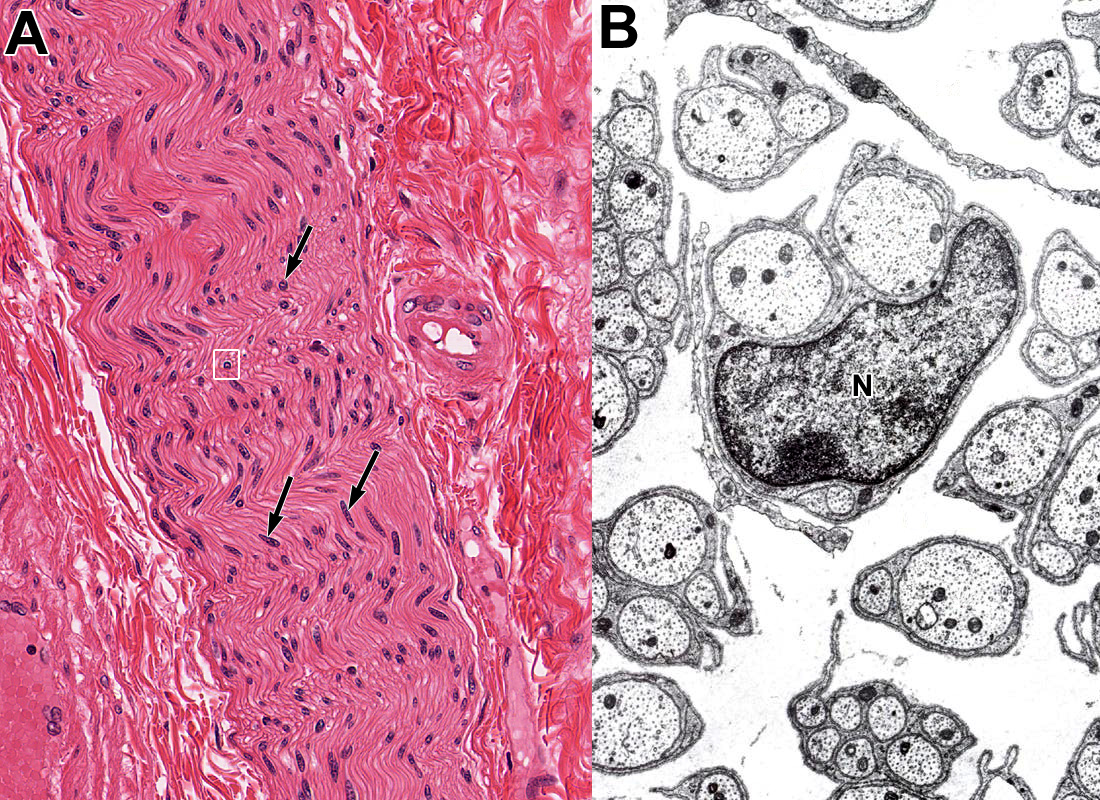
1. The nuclei indicated by the arrows in Panel A and labeled "N" in the electron micrograph in Panel B (which is a high magnification view of the boxed area in panel A) belong to which type of cell?
View Image
- Schwann cells
- Smooth muscle cells
- Fibroblasts of the perineurium
- Dorsal root (sensory) ganglion neurons
- Autonomic ganglion neurons
Answer
Correct answer 1. Schwann cells as the images depict a myelinated nerve in Panel A and an unmyelinated nerve in Panel B.
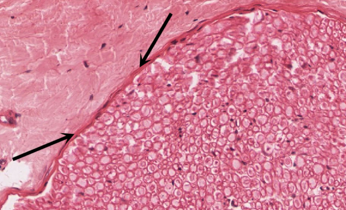
2. What type of cell junction plays an important role in the FUNCTION of the connective tissue layer indicated by the arrows?
View Image
- Gap junction
- Desmosome
- Hemidesmosome
- Zonula adherens or adherent junction
- Tight junction
Answer
Correct answer 5. Tight junctions in the perineurium.
3 Identify the cells indicated by the arrows in the below micrograph.
View Image
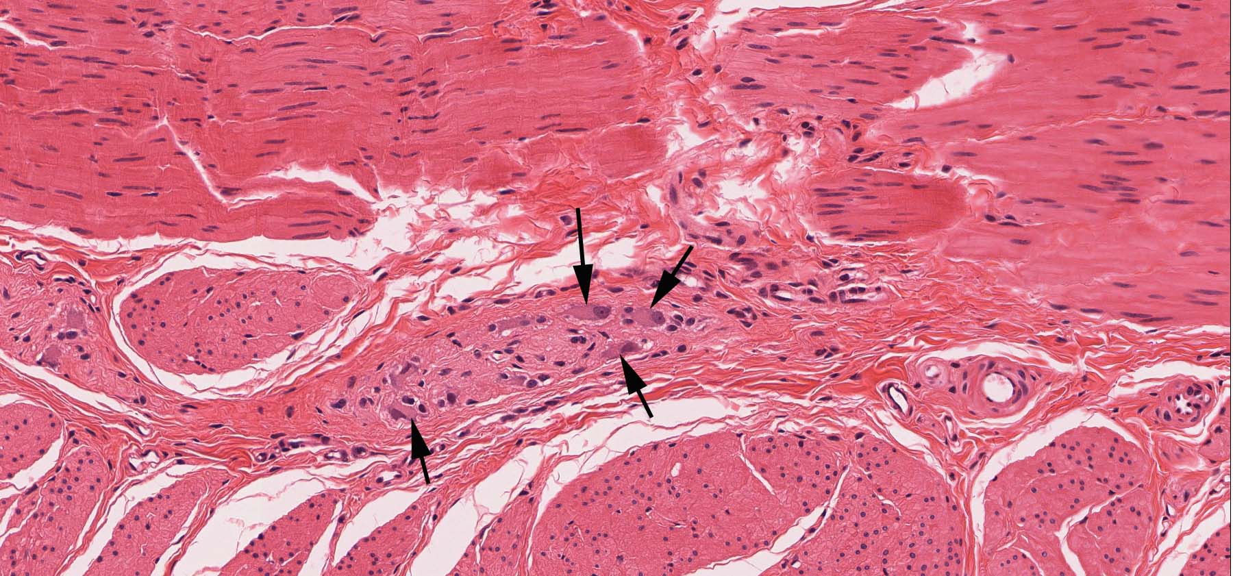
- Dorsal root ganglion neurons
- Autonomic ganglion neurons
- Satellite cells
- Schwann cells
- Intrafusal muscle fibers (cell)
Answer
Correct answer 2. Autonomic ganglion neuronal cell bodies between two layers of smooth muscle cells. The image depict a myenteric plexus or Auerbach's plexus.