- Be able to recognize compact and cancellous bone in conventional and ground sections and know the structural differences between the two types.
- Be able to identify the component parts of adult bone and know their functions (e.g. periosteum, endosteum, osteon, canaliculus, lacunae, osteocyte, Haversian systems/canals and Volkmann’s canals).
- Be able to recognize the cells in adult bone at the light and EM levels and know their functions (e.g. active and inactive osteoblasts, osteocytes, osteoclasts).
- Know the major differences in matrix contents of cartilage and bone.
- Know the location of the stem cells that are involved in making and repairing bone?
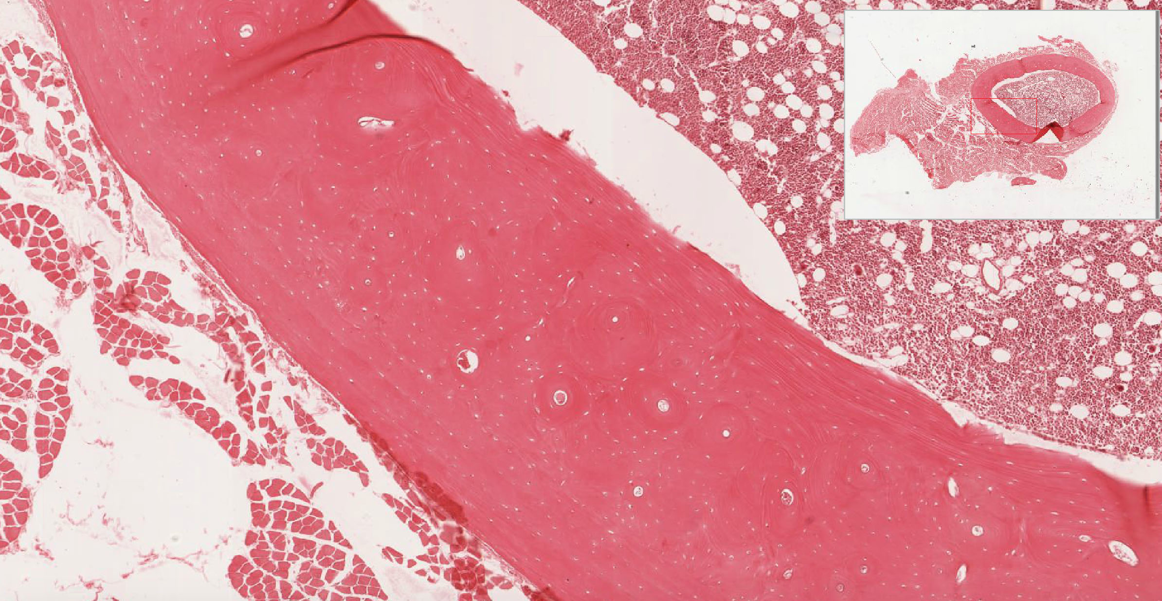
Slide 50 (fibula, monkey, decalcified, H&E) View Virtual Slide
Prior to sectioning and staining, this sample was soaked in a weak acid solution thus dissolving the mineralized component of the bone matrix but leaving behind all of the organic components (mostly type I collagen). Even though this section is distorted, you should be able to find osteons in various stages of development, lacunae, and canaliculi (to see canaliculi you will need to use your microscope and the glass slide from your collection --cut down the light by closing down the iris diaphragm to see them). The inner and outer circumferential lamellae slide 050 View Image can the bone shaft can also be seen in this section.
What distinguishes between compact and spongy bone?
Answer
Though it is easy to differentiate between compact and spongy bone at a gross level, at the microscopic level the main difference is the presence or absence of osteons. Grossly, compact bone has a dense appearance and is found, for example, on the outer surfaces of the long bones of the body. As the name implies, spongy bone is shaped like a sponge. The spaces within the sponge-shaped framework are filled with bone marrow. Compact bone, microscopically, is made of numerous osteons, whereas spongy bone is composed of sheets of lamellar bone and does not contain osteons.
Ground sections:
Cross sections:
Slide 051 Bone Ground section of compact cross View Virtual Slide
Slide 051XC Bone Ground section fibula cross View Virtual Slide
Slide 093B Bone Ground section of compact bone H&E cross section View Virtual Slide
Longitudinal sections:
Slide 093A Bone Ground section of compact longitudinal View Virtual Slide
Slide 051 Bone Ground section of compact longitudinal View Virtual Slide
Slide 051L-EX Bone Ground section of compact longitudinal View Virtual Slide
Slide 093C Bone Ground section of compact bone longitudinal View Virtual Slide
These "ground sections" were prepared by taking pieces of bone and grinding them with abrasives between two glass plates until they are thin enough to be semi-transparent. First, study cross sections (slides 51 and 93B). In these sections, the trapped air bends the light giving a dark image; the mineral and matrix generally transmit the light. You should be able to identify osteons and their subdivisions (as in slide 50), interstitial lamellae, Haversian/central canals and nutrient canals (Volkmann). Note that the latter canals penetrate osteons without causing new lamellae to be laid down around them. Note that slide 51XC is also an entire cross section of the fibula, so you should try to compare it against slide 50 discussed above.
Study the thinnest ground section (slide 93A) to identify lacunae and canaliculi. Now, look at the longitudinal sections (slides 51-20x, 51-40x, or 93C) of compact bone and try identifying the various structures mentioned above, especially Haversian/central and Volkmann's canals.
90 Osteocyte View Virtual EM Slide
The calcium crystals of the bone matrix were removed in this preparation by a decalcification process. Note how coarse the collagenous fibrils are and the difficulty in visualizing the periodicity of the fibrils (probably due to the process of mineralization).
93 Haversian canal View Virtual EM Slide
Note the "inactive" appearance of endosteal cells. The presence of a macrophage in the Haversian canal indicates the potential eroding function of the endosteal lining of the canal. Why are blood vessels so important in bone? (MatureBO2)
Click on a question to reveal the answer.
What distinguishes between compact and spongy bone?
Though it is easy to differentiate between compact and spongy bone at a gross level, at the microscopic level the main difference is the presence or absence of osteons. Grossly, compact bone has a dense appearance and is found, for example, on the outer surfaces of the long bones of the body. As the name implies, spongy bone is shaped like a sponge. The spaces within the sponge-shaped framework are filled with bone marrow. Compact bone, microscopically, is made of numerous osteons, whereas spongy bone is composed of sheets of lamellar bone and does not contain osteons.
Why are blood vessels important in bone?
Bone is a living tissue and, as such, needs a constant supply of nutrients. Without blood vessels, the bone would die.
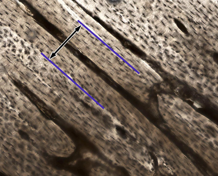
1. In this section of ground bone, identify the area indicated between the two purple lines.
View Image
- Outer circumferential lamellae
- Interstitial lamellae
- A Haversian system (osteon)
- A Volkmann's canal
- Inner circumferential lamellae
Answer
correct answer 3. A Haversian system (osteon) - this is a longitudinal section of compact bone showing LONGITUDINAL profiles of osteons consisting of concentric lamellae surrounding central Haversian canals (also in longitudinal profile). The canals that run PERPENDICULAR to the Haversian canals and it do NOT have any concentric lamellae organized around them are nutrient or Volkmann's canals.